Anatomy is not an easy subject, but it can be made easier with 3D interactive anatomy resources which makes organs and body parts come alive!
The TMC Library offers a variety of anatomy resources:
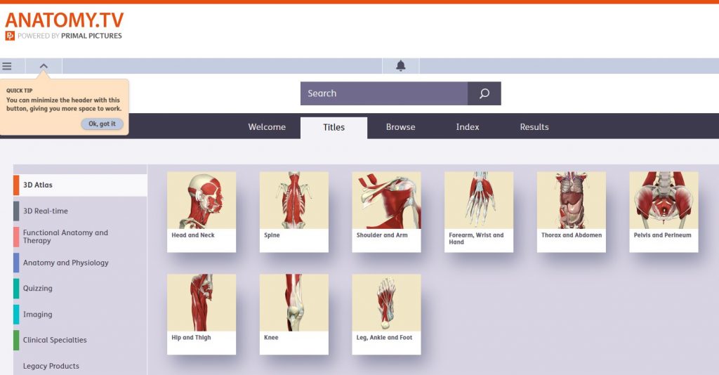
Anatomy TV, published by Primal Pictures, claims to be the world’s most medically accurate and detailed 3D graphic rendering of human anatomy based on imaging data. It is our most popular 3D anatomy tool. It offers animations which demonstrate functions, biomechanics, and surgical procedures. It offers quizzes and a mobile app with the ability to see labels in different languages, including Chinese and Latin. To supplement the core three-dimensional anatomy data are clinical videos and text written by leading medical specialists.
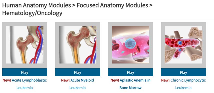
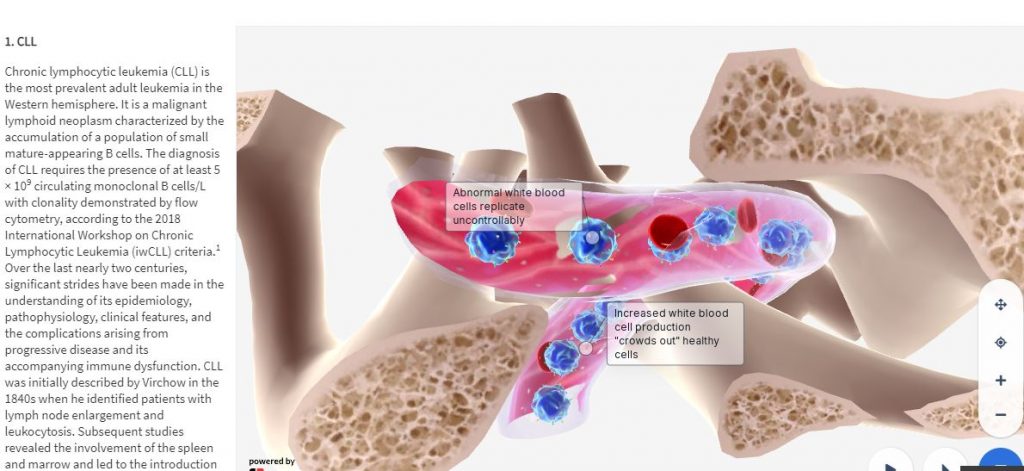
BioDigital Focused System Modules is a collection of over 300 interactive 3D modules that cover 17 specialties. These are not just anatomy modules, but simple animated illustrations of organs, diseases, processes, and conditions affecting the human body. Like Anatomy TV, many of its animations are also based on imaging data.
In addition to these subscription-based anatomy resources, there are several anatomy resources that are freely available online:
Get Body Smart: An Online Examination of Human Anatomy and Physiology is an application designed to “help explain the body’s complex physiological interactions and illustrate its important anatomical landmarks.” Diagrams and drawings as well as free quizzes and tutorials are included. General subject divisions include: skeletal system, muscle tissue physiology, muscular system, nervous system, circulatory system, respiratory system, urinary system, and histology. It contains quizzes to help students get test sharp.
IMAIOS: e-Anatomy, e-Cases, e-Courses This French resource provides a multi-unit online course on MRI (Magnetic Resonance Imaging) physics is fully open access. Intended for radiology residents, radiologists, MRI techs, medical physicists and students, its content includes interactive animations, experiments and quizzes designed to add fun while learning MRI physics. Not all of it is open access, however.
Netter Images is a collection of images by Frank Netter, M.D. and other excellent medical illustrators is the online version of the CIBA collection. Some collections can be browsed here, along with two texts: Netter’s Concise Atlas of Orthopaedic Anatomy and Netter’s Head and Neck Anatomy for Dentistry.
Neuroscience Online is interactive courseware for the study of neuroscience is provided by the Department of Neurobiology and Anatomy at The University of Texas Medical School at Houston. The project is being developed under the direction of the Department Chair and Editor, John H. Byrne. A companion resource to this textbook, Neuroanatomy Online, has been created in collaboration with Medical Neuroscience course director, Nachum Dafny.
Open Anatomy Browser The Open Anatomy Browser (OABrowser) is an open source, web-based anatomy atlas viewer. OABrowser displays three-dimensional anatomical models, image cross-sections of labeled structures and source radiological imaging, and a text-based hierarchy of structures. Included within it are several anatomy atlases (an MRI-derived brain atlas and atlases of other parts of the anatomy).
Whole Brain Atlas This is a neuroimaging educational tool showing trans-axial, sagittal and coronal views of the brain in cross-sectional MRI, CT and nuclear imaging techniques, with side-by-side navigation. Image series are provided for the normal brain and for its disease states: cardiovascular, neoplastic, degenerative and inflammatory/infectious. In disease states, clicking a link reading “clinical” will bring the user to a page of text explaining the case being viewed. The “Top 100 Brain Structures” feature is a study tool allowing the user to view structures individually, or to view a group of brain structures marked with pointers, labelled or unlabeled. The site is co-sponsored by the Departments of Radiology and Neurology at Brigham and Women’s Hospital, Harvard Medical School, its Countway Library of Medicine and the American Academy of Neurology.
Coming soon to The TMC Library are anatomy and physiology coloring books. Copy the pages and color them for relaxation and reinforcement.